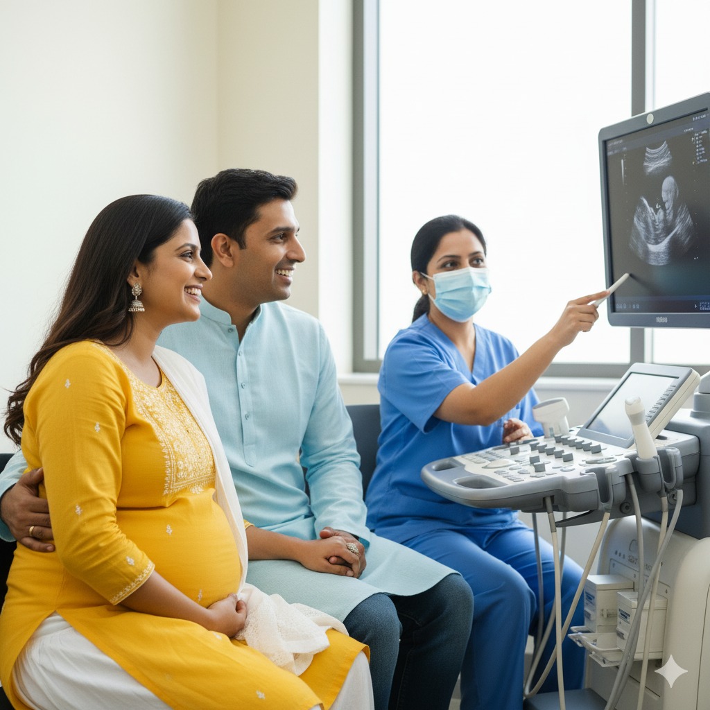
Fetal Echocardiography in kolkata
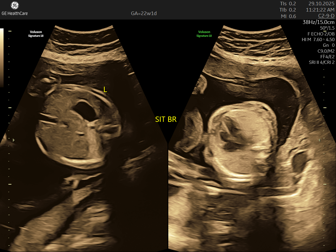
Fetal echocardiography is a valuabe early screening tool for Fetal Heart Defects. It is a specialized ultrasound scan that closely examines your baby’s heart while still in the womb. Unlike standard pregnancy scans, this test focuses on evaluating the structure, function, and rhythm of the fetal heart—providing incredibly detailed insights that help detect and diagnose congenital heart defects (CHDs) and other abnormalities early.
How is Fetal Echocardiography Different from Other Scans?
- Focus on the Heart: This scan is dedicated solely to assessing your baby’s heart—its structure, blood flow, and rhythm—offering a far deeper evaluation than standard ultrasounds.
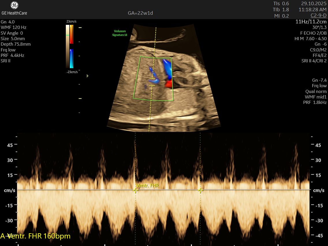
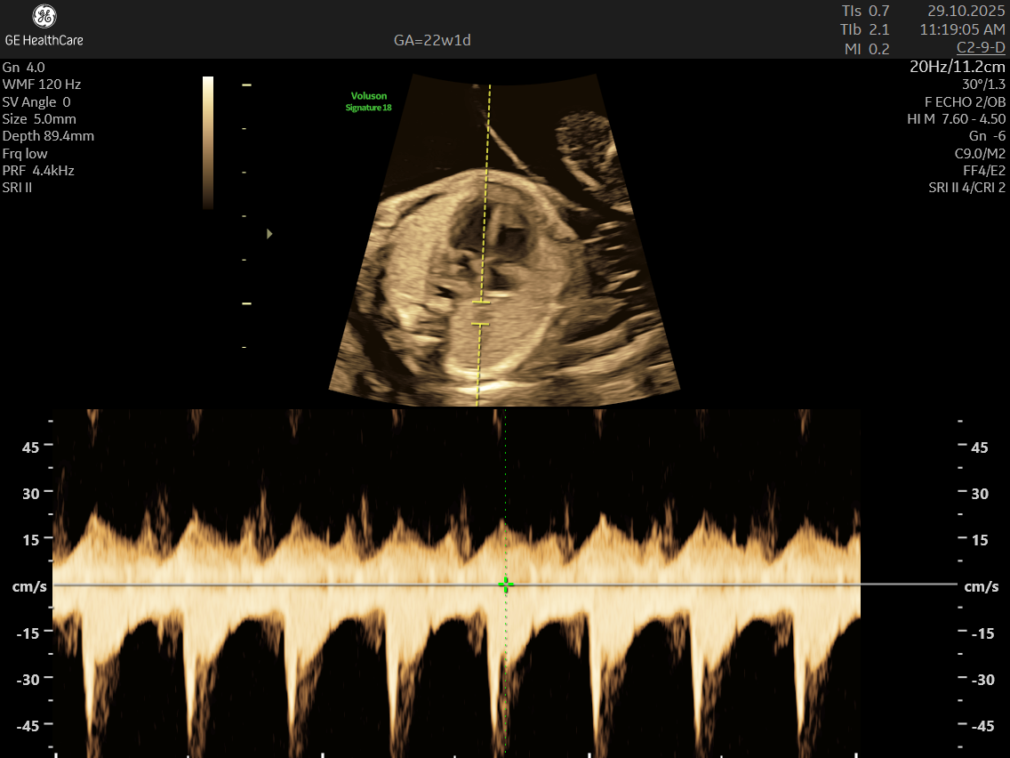
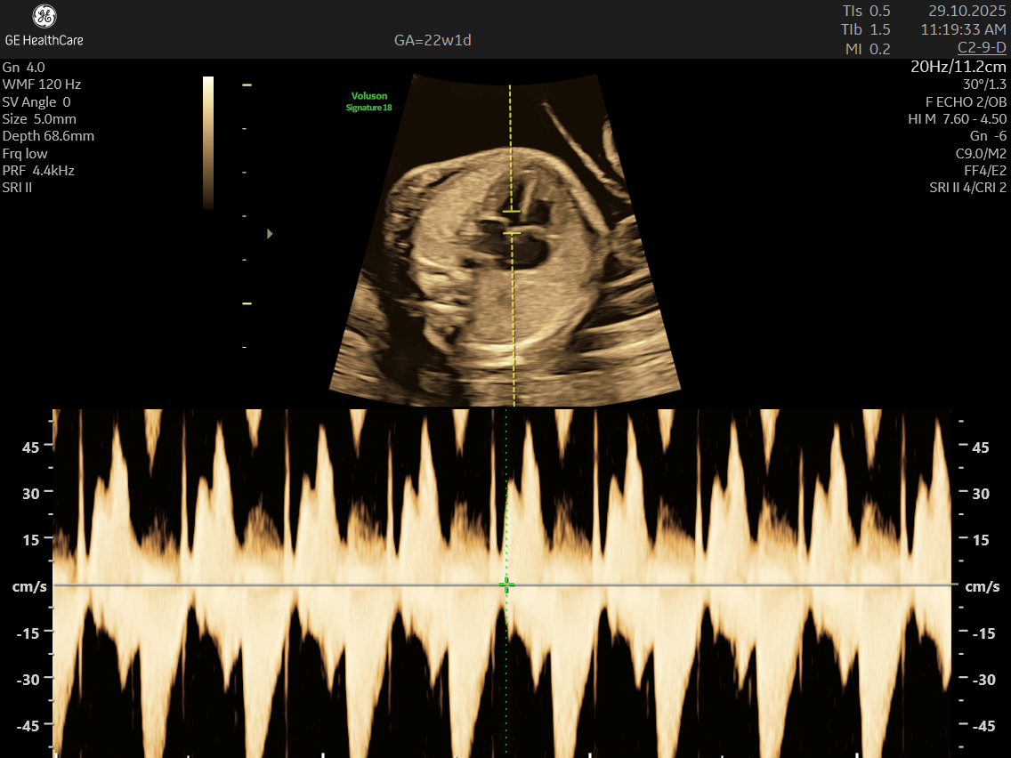
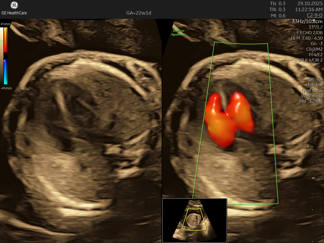
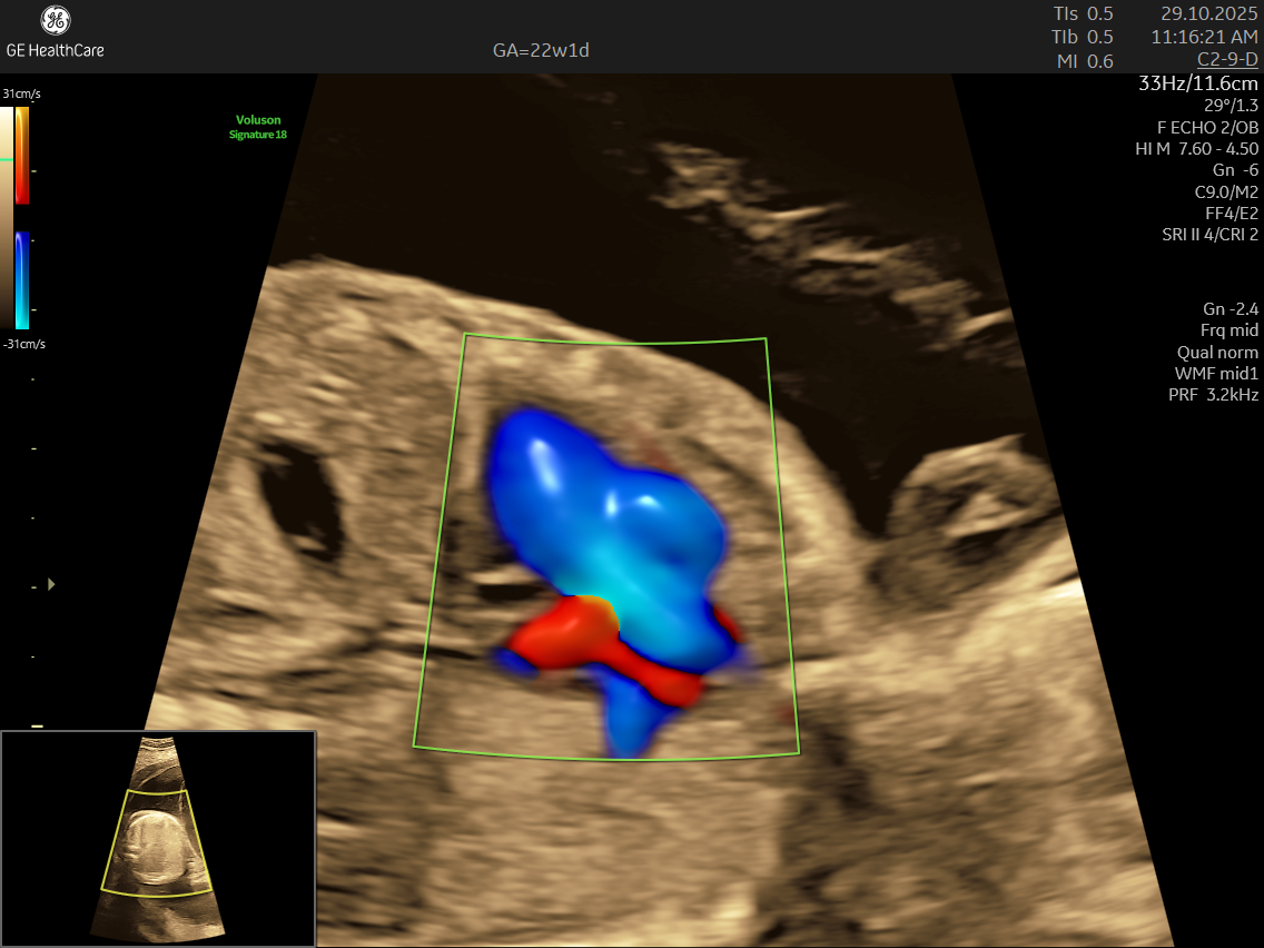
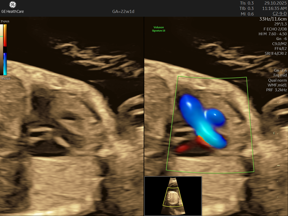
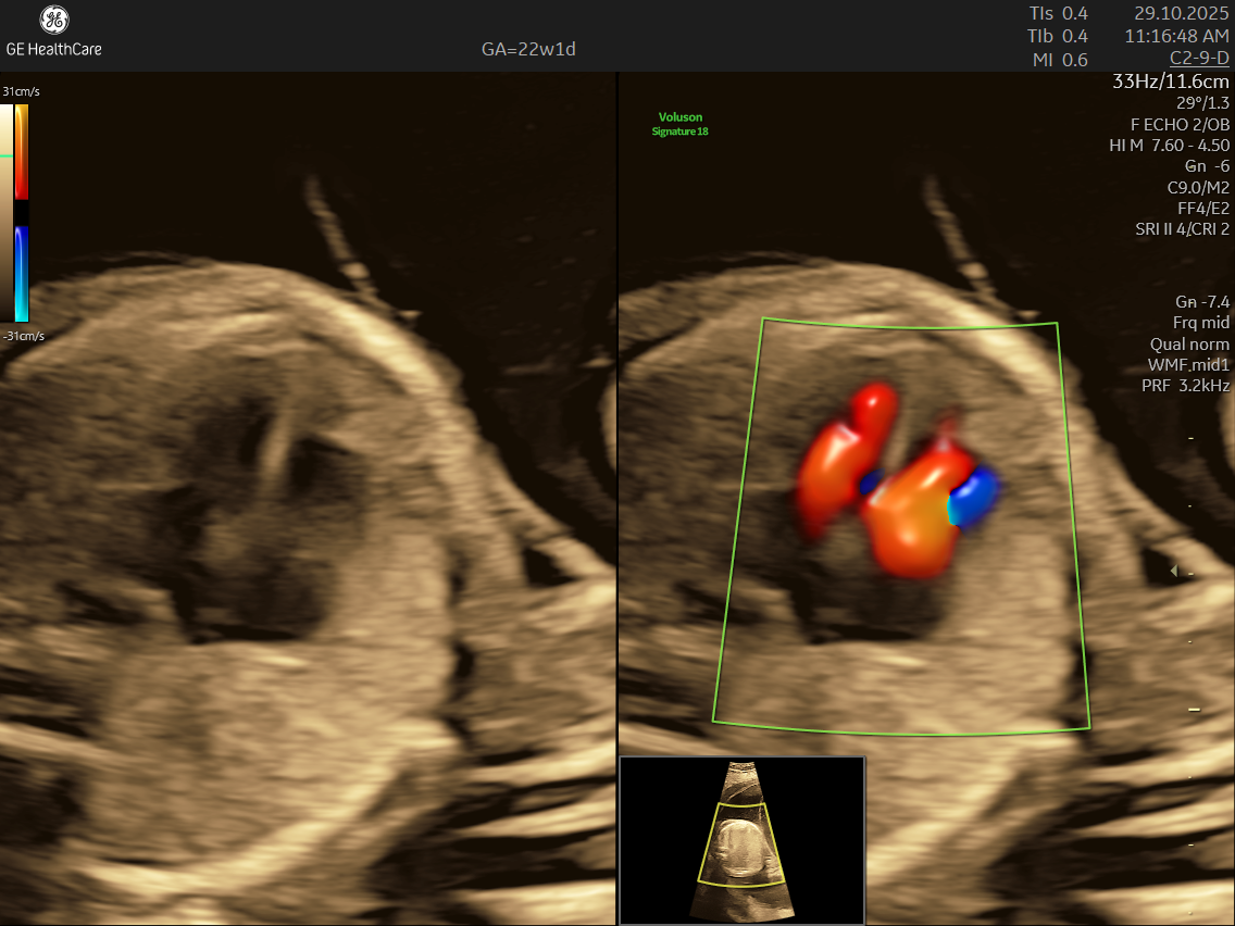
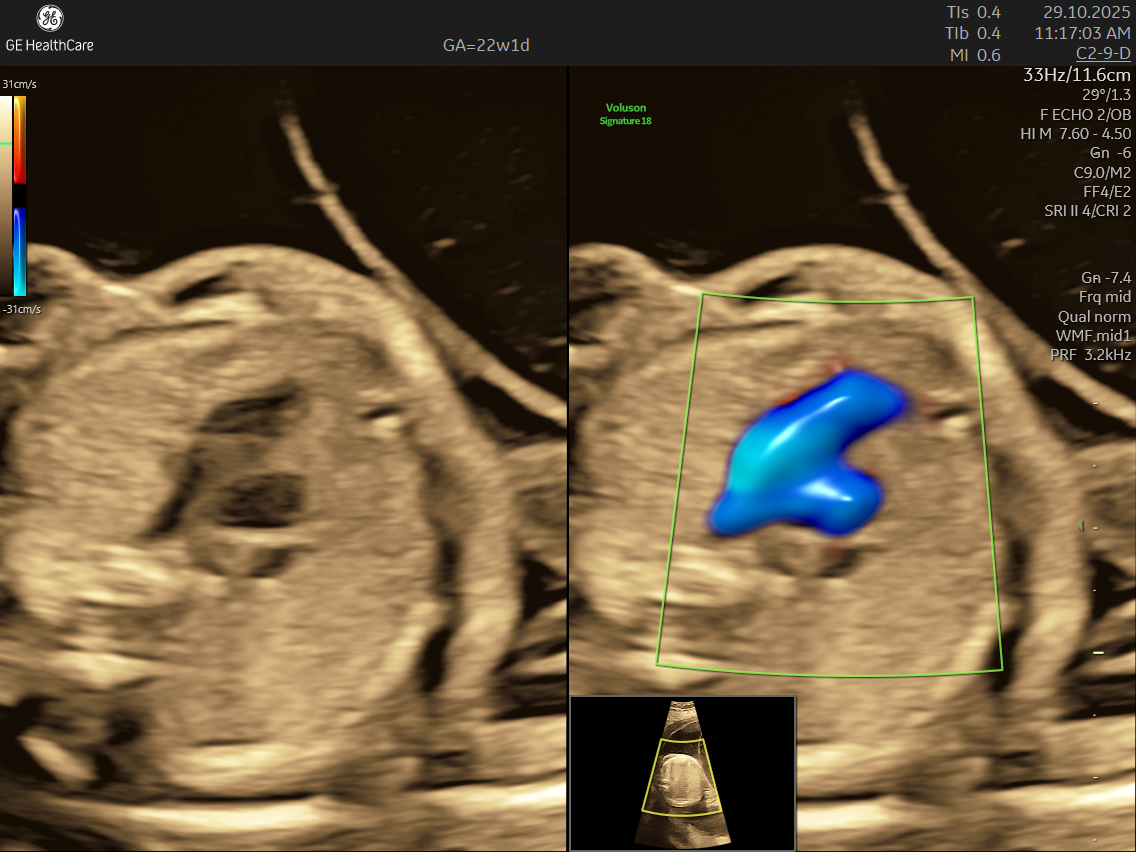
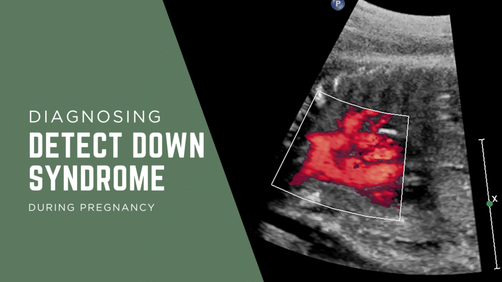
- High Detail: Utilizing advanced Doppler ultrasound technology, it tracks blood movement through the heart and valves, highlighting issues that general scans often cannot detect.
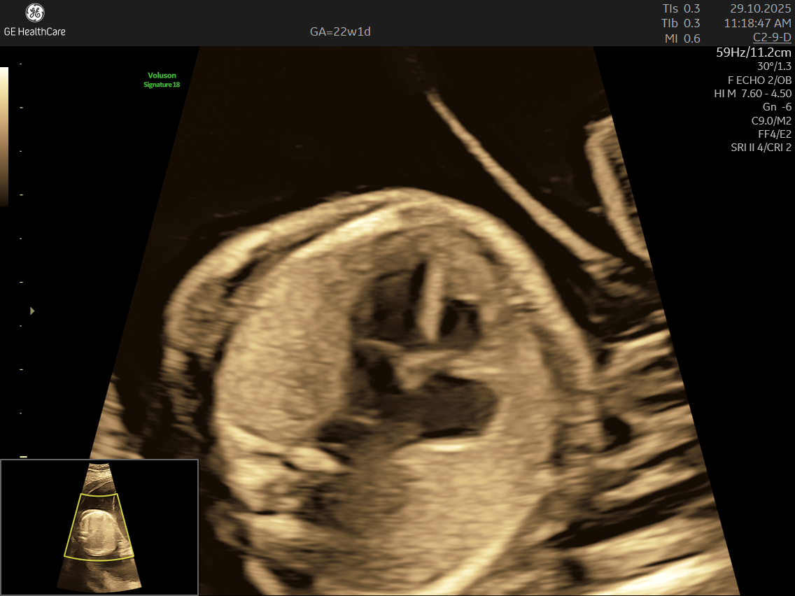
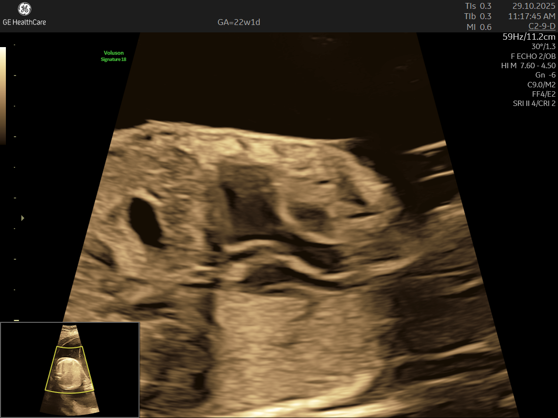
- Specialized Equipment and Expertise: Performed with high-resolution machines and by specialist doctors, fetal echocardiography pinpoints tiny structural or functional abnormalities that conventional scans may miss—helping ensure early, accurate diagnosis and informed planning.
When is fetal echocardiography recommended?
You should undergo fetal echocardiography if :
- You have a history of heart defects in the family or a previous child with CHD
- You have medical conditions such as diabetes or autoimmune diseases
- Routine ultrasound scans reveal abnormal heart rhythms or other concerns
- Pregnancy was achieved through assisted conception (IVF, ICSI)
- You were exposed to specific medications, infections, or substances that may impact fetal heart development
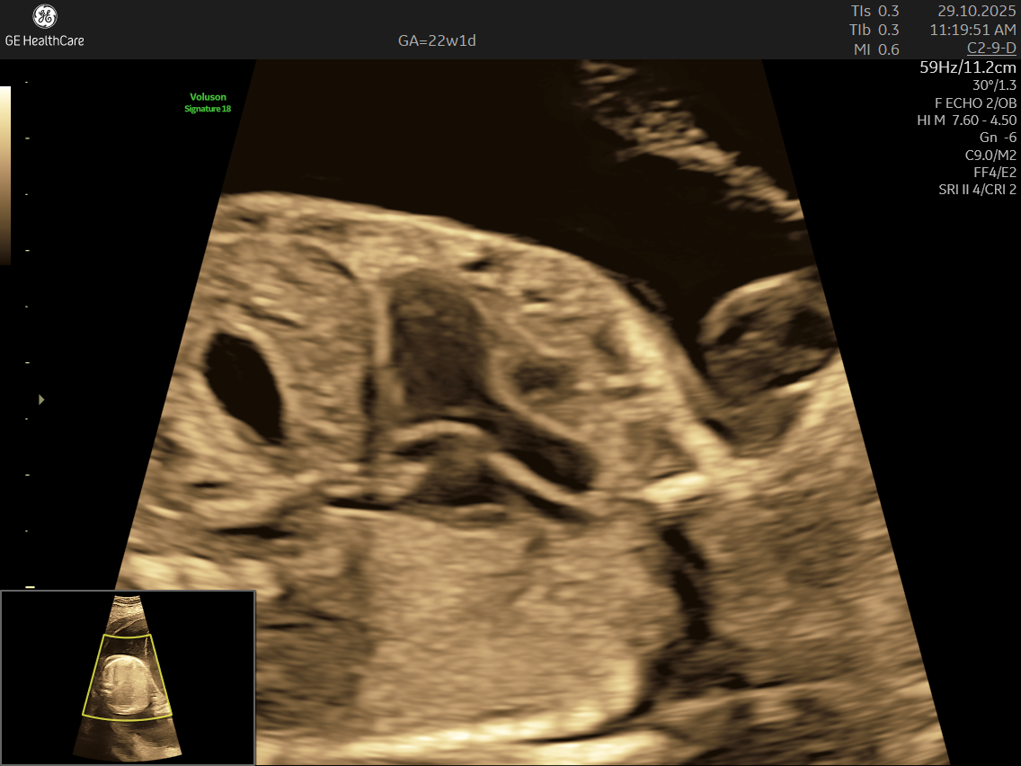
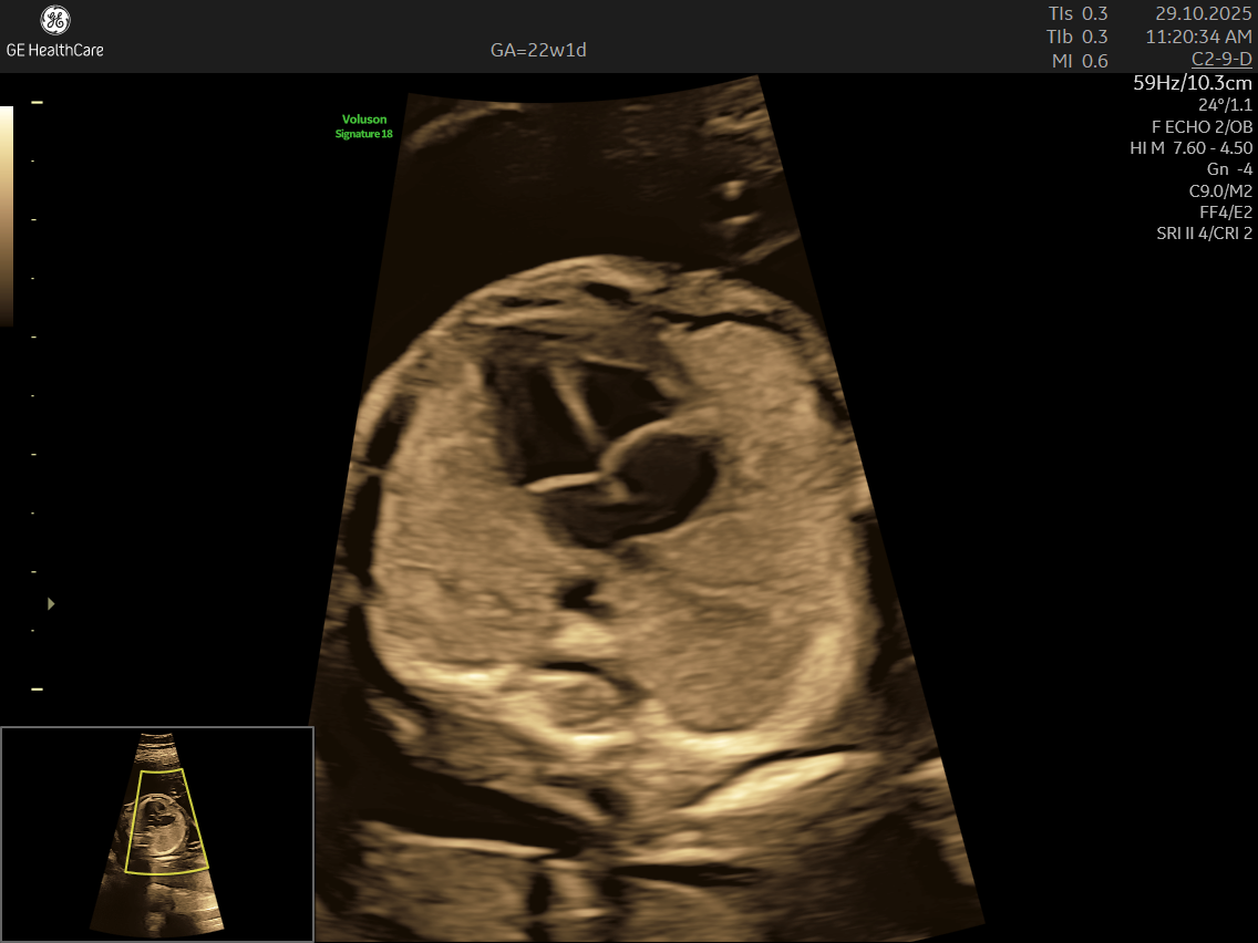
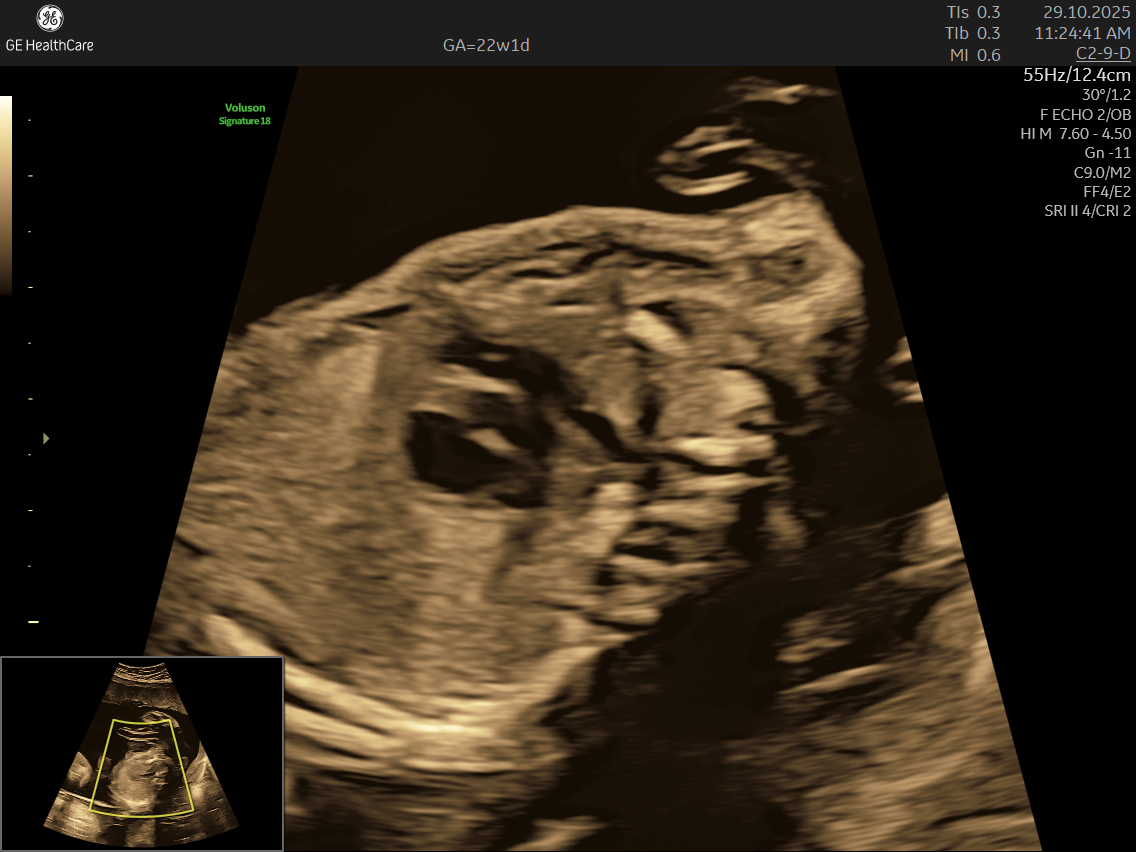
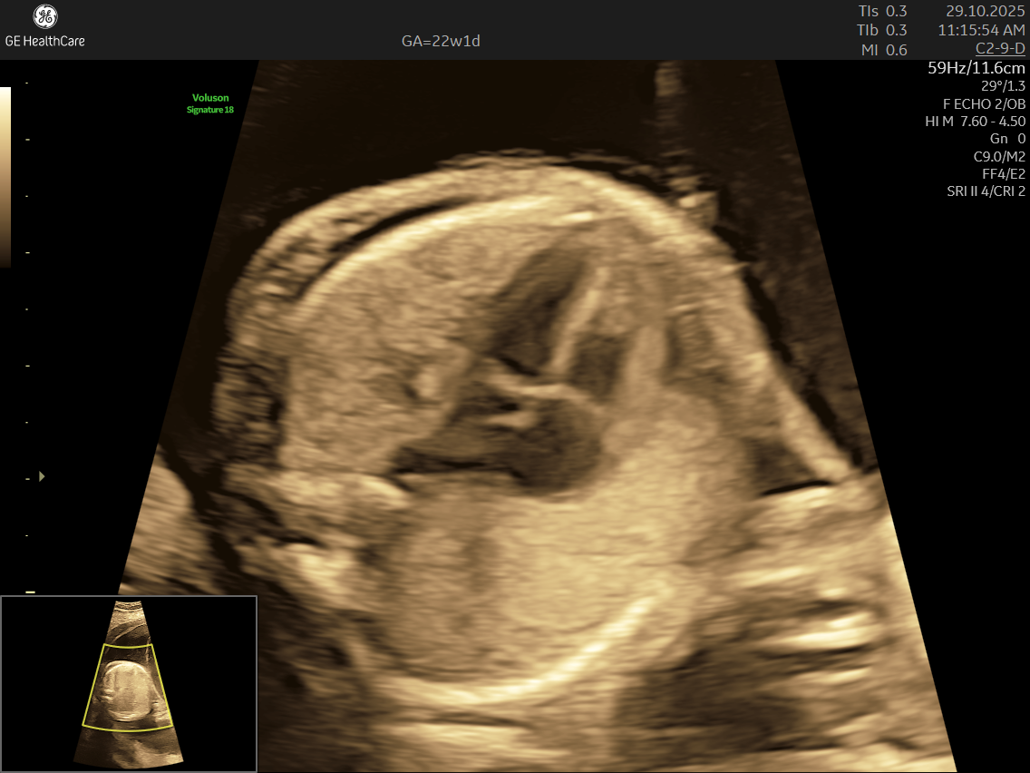
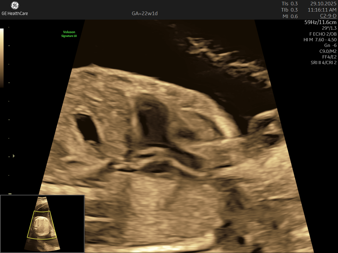
What Does the Scan Look For?
Fetal echocardiography examines:
- Structural Defects: Such as septal defects (holes in the heart), valve abnormalities, and underdeveloped chambers.
- Blood Flow Patterns: Detects arrhythmias or irregular heartbeats and checks for efficient circulation through the heart and major vessels.
- Rhythm Abnormalities: Detects irregular heartbeats or arrhythmias.
- Functional Issues: Identifies problems with the heart’s pumping efficiency.
- Syndromic associations: Heart issues linked to genetic syndromes (like Down or Turner syndrome)
Who can perform a Fetal Echocardiography?
Fetal echocardiography is typically performed by a fetal medicine specialist, pediatric cardiologist, or a specialist trained in fetal echocardiography because:
- Congenital heart defects are complex and require expertise for accurate interpretation.
- Identifying heart abnormalities demands a deep understanding of embryology, cardiac anatomy, and physiology.
- Specialists ensure precise evaluation to guide further interventions or treatment plans.
What are the Abnormalities that can be detected by Fetal Echocardiography?
Fetal Echocardiography can detect a wide range of abnormalities in the fetal heart, including congenital heart defects, arrhythmias, and structural anomalies. Some of the key abnormalities detected by fetal echocardiography are:
- Ventricular septal defects (VSD), pulmonary stenosis, right ventricular hypertrophy, and overriding aorta (Tetralogy of Fallot).
- Transposition of the great arteries, where the aorta and pulmonary artery are switched.
- Hypoplastic left heart syndrome, involving underdevelopment of the left side of the heart including the left ventricle, mitral valve, aortic valve, and aorta.
- Common atrioventricular canal defects involving large holes at the center of the heart affecting atrial and ventricular septa along with AV valves.
- Coarctation of the aorta, seen as narrowing of the aorta near the ductus arteriosus.
- Arrhythmias like premature atrial and ventricular contractions, supraventricular tachycardia, and complete heart block.
- Echogenic intracardiac foci, which can be benign but sometimes associated with chromosomal anomalies such as trisomy 13 or 21.
- Malformations of main arteries (aorta and pulmonary artery), underdeveloped heart chambers, abnormalities of valves (pulmonary, aortic, tricuspid).
- Abnormalities in blood flow patterns and valve function observed on Doppler imaging.
Why choose NESA Insitute of Fetal Medicine for Fetal Echocardiography?
At NESA Insitute of Fetal Medicine, expectant couples receive their scan:
- In a comfortable, hygienic environment designed to minimize stress and ensure safety during your scan.
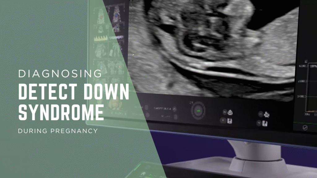
- Using the advanced GE Voluson S10 ultrasound machine—renowned for its superior imaging clarity, 4D real-time visualization, and automated analysis tools that significantly increase diagnostic accuracy and reduce the chances of missing subtle heart defects.
- Under the care of experienced fetal medicine panel of experts lead by Dr Khurshid Alam, Kolkata’s Leading fetal medicine Specialist who take time for detailed, thorough examinations and provide trustworthy, actionable reports.
- Its a specialized ultrasound done between 18–24 weeks of Gestational Age that evaluates the structure, function, and rhythm of a baby’s heart while still in the womb. This scan uses high-frequency sound waves to create detailed images of the fetal heart, aiding in the detection of congenital heart defects (CHDs) and other abnormalities.
- Since interpreting fetal heart scans requires specialist knowledge and experience, your scan will be performed and reported by accredited professionals in fetal medicine or pediatric cardiology. NESA is recognized across Kolkata for its specialist team and commitment to maternal and fetal health.