Fetal Anomaly Scan in Kolkata
Looking for a reliable and trusted Fetal Ultrasound centre for a Fetal Anatomy scan in Kolkata? At NESA Institute of Fetal Medicine, you can get your scan done by Kolkata’s most trusted Fetal Medicine Specialists.
What to expect at your Detailed Fetal Anatomy Scan at NESA Institute of Fetal Medicine
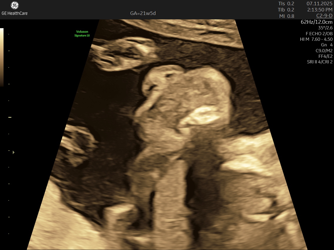
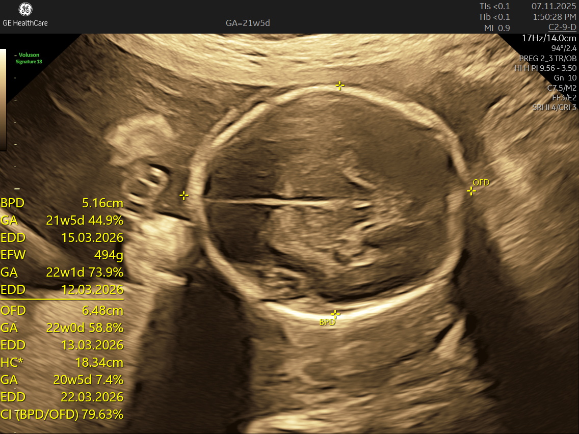
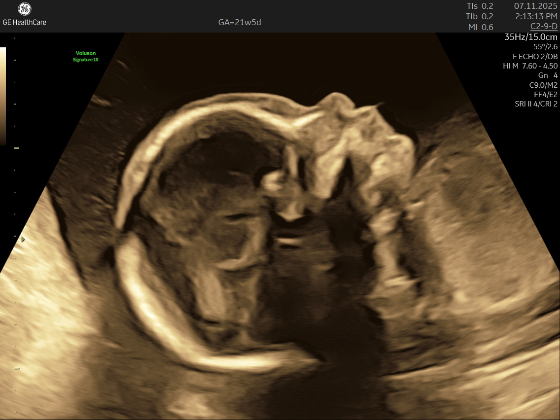
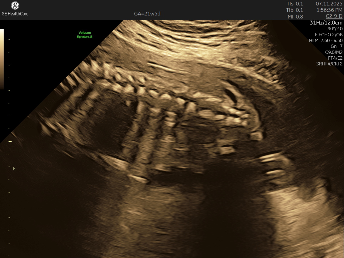
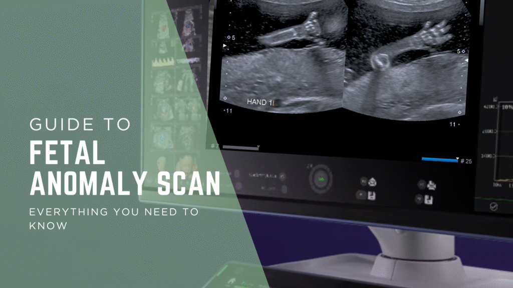
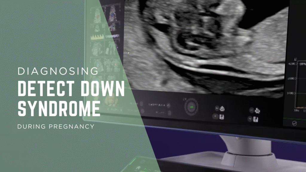
If you’re expecting a baby, the Detailed Anatomy Scan —also known as the Fetal Anomaly Scan — is one of the most important checks you’ll have during pregnancy. Usually done between 18 and 22 weeks, this ultrasound is performed by highly trained fetal medicine specialists at NESA Institute of Fetal Medicine. The scan provides a close-up look at your baby’s overall development and helps to make sure everything is progressing as it should.
During this scan, the MFM specialist carries out a detailed examination of your baby’s body, checking their vital organs, spine, head, heart, limbs, and more. The aim is to confirm that your baby’s growth is on track and can identify various structural abnormalities and potential complications early, so that appropriate steps can be taken early.
Why Is a Fetal Anomaly Scan So Important?
For parents, the reassurance that baby is healthy and thriving can relieve a lot of anxiety. If the scan picks up any concerns, such as congenital heart defects or neural tube issues, your care team can take action right away. This might include expert consultations, additional scans, or, in rare cases, planning for treatment after birth.
Early detection means you get:
Peace of mind: Most scans confirm healthy development and offer comfort for the rest of your pregnancy journey.
Expert support: Prompt guidance from specialists to help you understand findings and make confident choices.
Better outcomes: If needed, care plans or treatments can be discussed and arranged before your baby is born.
What Does the Fetal Medicine Specialist Check During a Fetal Anomaly Scan?
This scan provides reassurance by confirming your baby’s major organs and bones are developing properly. If anything unusual is detected, your doctor will guide you on the next steps and offer comprehensive support throughout. The specialist will check for:
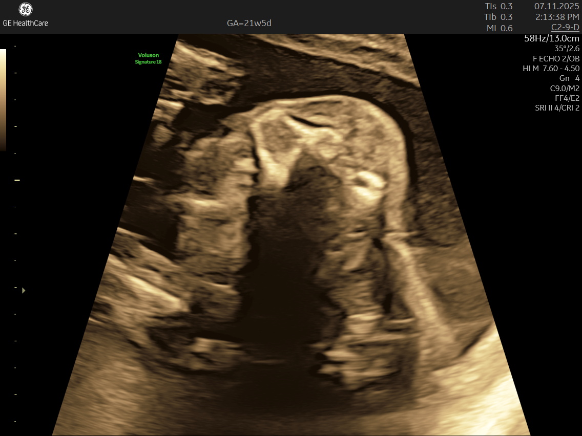
- Head and Brain: The scan looks at the baby’s skull shape and examines the brain’s key areas, checks the structure and growth of the brain and skull, ensuring everything is forming correctly.
- Face: Checks the eyes, lips, and nose, ruling out conditions like cleft lip/ palate.
- Head and Brain: The scan looks at the baby’s skull shape and examines the brain’s key areas, checks the structure and growth of the brain and skull, ensuring everything is forming correctly.
- Face: heck the eyes, lips, and nose, ruling out conditions like cleft lip.
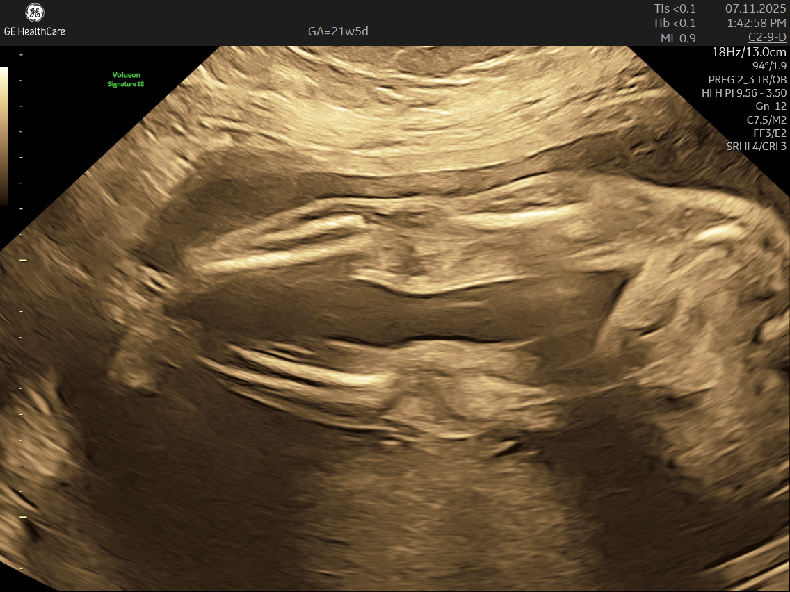
- Spine and Back: Assesses alignment and formation. A healthy, well-formed spine is a crucial marker of baby’s overall development.
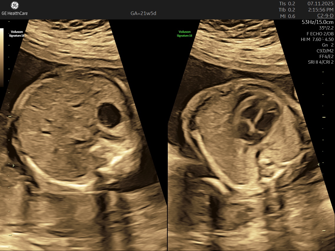
- Heart: The scan takes time to review the heart’s structure and rhythm, checking for chambers, valves, and major blood vessels; screens for heart defects
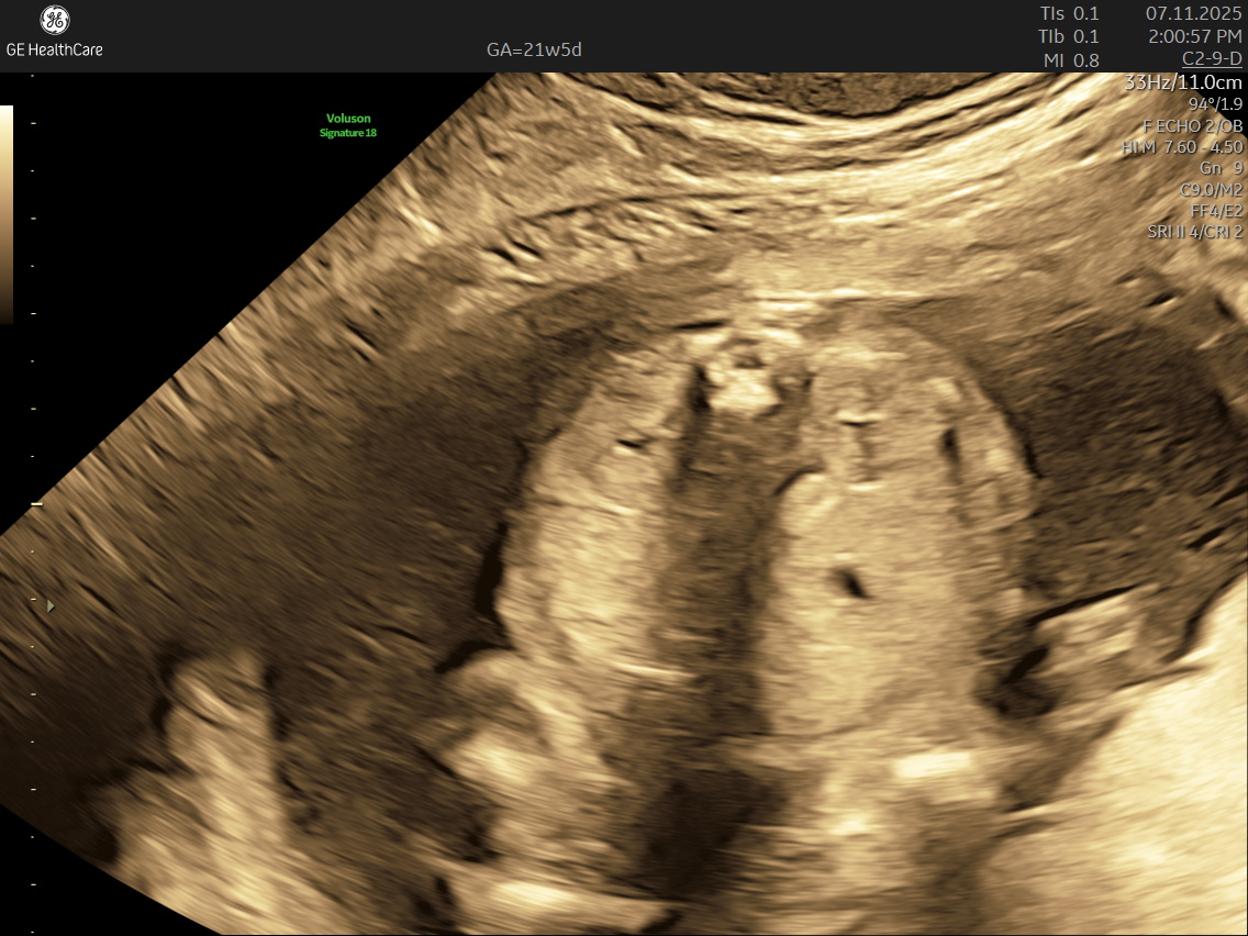
- Abdomen and Organs: Sonographers measure your baby’s tummy, assess the position of important organs like the stomach, kidneys, bladder and intestines.
- Limbs: Each arm and leg is carefully viewed to make sure they’re growing and moving as expected.
- Placenta and Amniotic Fluid: Checks that your baby’s environment is safe, and the placenta is in the right place to nourish your baby.
- Spine and Back: Assesses alignment and formation. A healthy, well-formed spine is a crucial marker of baby’s overall development.
- Heart: The scan takes time to review the heart’s structure and rhythm, checking for chambers, valves, and major blood vessels.
- Abdomen and Organs: Sonographers measure your baby’s tummy, assess the position of important organs like the stomach, kidneys, and bladder.
- Limbs: Each arm and leg is carefully viewed to make sure they’re growing and moving as expected.
- Placenta and Amniotic Fluid: Checks that your baby’s environment is safe, and the placenta is in the right place to nourish your baby.
Which Conditions Can Be Detected in the Fetal Anomaly Scan?
- The scan is detailed and covers a wide range of possible conditions:
- Neural tube defects like spina bifida.
- Facial differences (cleft lip/palate).
- Heart defects (congenital problems, valve issues).
- Abdominal wall defects (such as an opening where organs may protrude).
- Limb concerns, including clubfoot.
- Kidney and urinary issues, sometimes spotted if kidneys or bladder aren’t developing properly.
- Growth challenges such as too little or too much growth.
- Placental issues, like placenta previa.
- Genetic or chromosomal markers, supporting further testing if needed.
What Happens If Something Is Found During the Scan?
Most babies are found to be healthy, but in the event the scan raises questions, you’re not alone—your medical team will walk you through the next steps. If the scan shows a potential issue, it does not always mean something is wrong. If the Detailed Fetal Anatomy (Anomaly) Scan finds that your baby may have signs of a condition, here’s what typically happens:
- Follow-Up: Regular monitoring, additional scans, and continuous guidance will be provided to track your baby’s development and prepare you for delivery and newborn care.
- Further Evaluation: Your doctor will explain the findings and may recommend repeat or specialized ultrasounds to confirm the results.
- Diagnostic Tests: Depending on the suspected issue, tests such as fetal echocardiography (for heart concerns), genetic testing, or amniocentesis may be suggested to gather more detailed information.
- Specialist Consultations: You may be referred to fetal medicine experts, pediatricians, genetic counselors, and neonatologists who explain the condition, prognosis, and possible treatments in detail.
- Treatment Planning: The medical team will discuss management options, which may include planning for treatment before or after birth, adjusting your delivery plan, or providing specific interventions if needed.
- Decision Support: You’ll receive counseling and emotional support to help you understand your choices and make informed decisions for your baby and family
The caring team at NESA Institute of Fetal Medicine will ensure you understand every finding and throughout, the goal is to provide clarity, support, and the best possible care for both you and your baby. A specialised fetal Medicine clinic like NESA Institute of Fetal Medicine has all the specialist and technical expertise to ensure you are not alone—specialists help answer questions and guide your next steps with compassion and expertise. We support parents with clear information and compassionate guidance, helping you make informed decisions at every step.
Why Trust NESA Institute of Fetal Medicine in Kolkata?
Give your baby the best start – choose a Detailed Fetal Anatomy Scan in Kolkata at NESA Institute of Fetal Medicine. Here’s why:
- Trusted by new parents across Kolkata
- Top fetal medicine & radiology experts
- Cutting-edge ultrasound technology
- Personalized family-centric care
- Clean, comforting clinic environment